
1 in 2 May Be Infected. So How Do You Know If You Have H. pylori?
In China, the prevalence of Helicobacter pylori (H. pylori, or Hp) infection is as high as 40%–50%, meaning that nearly one in two people may be infected.
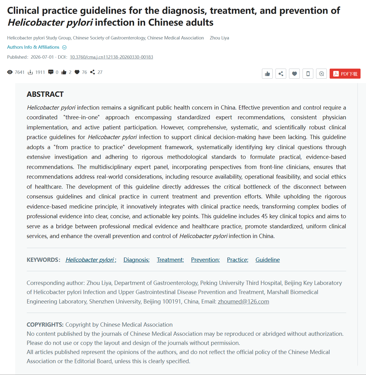
H. pylori infection is a major cause of chronic gastritis, peptic ulcers, and gastric cancer, making accurate diagnosis the first step toward effective management. In July 2026, the Helicobacter pylori Study Group of the Chinese Society of Gastroenterology released the latest edition of the Clinical Practice Guideline for the Diagnosis, Treatment, and Prevention of Helicobacter pylori Infection in Chinese Adults.
Based on a nationwide survey of 9,283 clinicians, the guideline addresses several of the most important questions surrounding diagnosis.
01 Symptoms Can’t Tell You Whether You’re Infected
Common misconception: Many people believe that “an upset stomach means you’re infected with H. pylori, and no symptoms means everything is fine.” This is actually a common misconception.
The guideline clearly points out that about 70% of people infected with H. pylori have no symptoms or signs at all. Even when symptoms do appear, they tend to be nonspecific-upper abdominal pain, bloating, feeling full after meals, early satiety, bad breath, and so on. These issues are not unique to H. pylori infection; chronic gastritis and functional dyspepsia can all produce similar symptoms.
More importantly, whether you have symptoms has no clear correlation with the severity of gastric mucosal damage, the future risk of developing gastric cancer, or the success of eradication therapy. In other words, a person who feels “perfectly fine” may already have gastric mucosal atrophy, intestinal metaplasia, or even precancerous lesions, while someone with “obvious symptoms” may actually have relatively mild mucosal damage.
Key message from the guideline: You cannot rely on the presence or absence of clinical symptoms to determine whether you are infected with H. pylori, whether to start eradication therapy, whether treatment has worked, or what the prognosis will be. The only way to know if you are infected is to get tested.
02 The Five Testing Methods: How to Choose the Right One?
The guideline systematically reviews the five main methods for diagnosing active H. pylori infection (that is, an infection currently present in the stomach) and compares their characteristics in detail. In simple terms, they fall into two broad categories: those that don’t require an endoscopy, and those that do.
2.1 Urea breath test — just breathe, and you’ll know
This is currently the most commonly used test and the first-choice follow-up method recommended by the guideline. The principle is clever: H. pylori produces a special enzyme called urease, which breaks down urea into carbon dioxide. During the test, you drink a small cup of urea solution labeled with a special isotope. If H. pylori is present in your stomach, it breaks down the urea and produces labeled carbon dioxide, which travels through the bloodstream to the lungs and is exhaled. By detecting the labeled carbon dioxide in your breath, doctors can tell whether you are infected.
The breath test comes in two versions: C13 and C14. C13 uses a stable isotope with no radioactivity and is suitable for everyone; C14 contains trace amounts of radiation (low energy and weak penetration) and is less expensive, but it is not recommended for pregnant or breastfeeding women and children. The two are equally accurate; which one to choose depends mainly on the equipment available locally and individual circumstances.
2.2 Stool Antigen and Nucleic Acid Tests — Infection Can Also Be Detected in Stool
Gastric mucosa colonized by H. pylori constantly sheds and passes through the intestines into the stool. The stool antigen test uses monoclonal antibodies to look for traces of H. pylori in stool, while the nucleic acid test goes a step further by directly detecting fragments of H. pylori’s DNA. Both methods are highly accurate, are unaffected by medications such as proton pump inhibitors (PPIs), and are not affected by the uneven distribution of bacteria in the stomach, making them excellent non-invasive options. The only minor “inconvenience” is that they require a stool sample, and some people may be less willing to cooperate.
2.3 Rapid urease test — a “one-minute test” during gastroscopy
During gastroscopy, the doctor takes a small tissue sample from the gastric mucosa and places it in a reagent containing phenol red and urea. If H. pylori is present, its urease breaks down urea to produce ammonia, raising the solution’s pH and turning its color from yellow to red. This method is simple to perform, yields rapid results and is low-cost, yet it can only be carried out during gastroscopy and is vulnerable to influences from biopsy sites and pre-test medications.
2.4 Gastric mucosal histology staining — “seeing it directly” under a microscope
Like the preceding test, a biopsy specimen is obtained during gastroscopy; however, in this case the tissue is sectioned, stained, and examined directly under a microscope for H. pylori.This method has very high specificity but relatively low sensitivity (it may miss the H. pylori). It is time-consuming and technically demanding. Its advantage lies in its ability to simultaneously assess pathological changes in the gastric mucosa, such as atrophy, intestinal metaplasia, and intraepithelial neoplasia.
Summary: If you are not undergoing gastroscopy, choose the breath test or stool test; if you are, a rapid urease test or histologic staining can be performed concurrently.
03 Does a Higher Breath Test Value Mean a More Severe Infection? Not Necessarily.
Many patients become anxious when they see a high value on their breath test report, and dismissive when the value is low. The guideline specifically addresses this misconception.
The breath test is essentially a qualitative test — it only tells you whether infection is present or absent. The actual value is influenced by many factors, including the number and density of bacteria, the urease activity of the strain, gastric emptying speed, the performance of the testing equipment, and how strictly the procedure is followed. It does not reflect the virulence of the bacteria or the severity of gastric mucosal damage, nor is it related to clinical outcomes or prognosis.
Worried when the value is high? Reassured when it’s low? Neither is correct. Positive is positive and negative is negative. The magnitude of the value only relates to diagnostic reliability — the further it is from the cutoff, the more reliable the result. When the value is close to the cutoff (for example, a C13 breath test value between 2 and 6), it is recommended to rule out interfering factors and retest, or to confirm with another method.
04 How to Follow Up After Treatment? Timing and Method Both Matter
H. pylori eradication therapy can fail. The guideline stresses that all patients who undergo eradication therapy must be re-tested. However, follow-up testing cannot be done at just any time or with just any method; there are three key points to keep in mind.
4.1 Timing: wait at least 4 weeks
You must wait at least 4 weeks after finishing eradication therapy before re-testing. Why can’t you test right away? Because the treatment drugs (especially antibiotics and acid-suppressive medications) suppress bacterial activity, which can lead to false-negative results. The bacteria need time to recover — if any remain, they will become active again after 4 weeks and can then be detected. The guideline recommends completing the re-test within 12 weeks and not waiting too long.
4.2 Method: the breath test is the first choice
The urea breath test is the first-choice follow-up method — it is simple, accurate, and unaffected by the focal distribution of bacteria in the stomach. Stool antigen or nucleic acid testing can serve as alternatives. A repeat gastroscopy is usually unnecessary, but if you have gastric mucosal lesions that require a follow-up gastroscopy, a rapid urease test or histologic staining can be done at the same time. A special note: serum antibody testing cannot be used to determine whether eradication was successful, because antibodies remain positive for a long time after eradication.
4.3 Medication: you must “abstain” before re-testing
Before re-testing, you need to stop medications that may interfere with the results: antibiotics and bismuth should be stopped for at least 4 weeks, and proton pump inhibitors (PPIs) and potassium-competitive acid blockers (P-CABs) for at least 2 weeks. These drugs suppress bacterial activity or alter the gastric environment, leading to inaccurate results.
05 A Positive Antibody Blood Test = Still Infected? Not Necessarily
Serum anti-H. pylori IgG antibody testing is one of the least expensive options — a single blood draw is all it takes, and it is unaffected by medications or gastric conditions. But it has one key limitation: a positive antibody result cannot distinguish between a current infection and a past infection that has already cleared.
After H. pylori infection, IgG antibodies gradually rise and remain elevated for a long time even after the infection clears. In other words, even if you have successfully eradicated H. pylori, your antibodies may still be positive. The guideline therefore clearly states that, except in special circumstances, IgG antibody testing cannot be used to diagnose active infection, justify starting treatment, or confirm eradication. A negative antibody result has some value in ruling out infection, but a positive result cannot be used as the basis for starting treatment.
However, in certain special situations, serum antibody testing still has unique value. For example, in patients with peptic ulcer complicated by acute bleeding, severe atrophic gastritis, status post partial gastrectomy, gastric mucosa-associated lymphoid tissue (MALT) lymphoma, or those who have difficulty stopping PPIs, other testing methods have high false-negative rates. For these patients, if they have never undergone eradication therapy, a positive serum IgG antibody result can serve as the basis for diagnosing active infection and starting treatment.
06 These “Little-Known Facts” You May Not Know
6.1 Drug resistance testing: making treatment more precise
Antibiotic resistance is the leading cause of H. pylori eradication failure. The guideline introduces two methods for resistance testing. The first is traditional culture-based phenotypic resistance testing, which can test multiple antibiotics but requires endoscopic sampling and is technically complex, with a low success rate and long turnaround time. The second is genotypic resistance testing, which detects resistance by identifying mutations in resistance genes; it can be performed on gastric mucosa, gastric juice, or even stool samples, and is simple and fast, with stool samples being particularly well accepted. Currently, genotypic testing performs well for detecting resistance to clarithromycin and levofloxacin, and relevant test kits have already been approved in China.
6.2 At-home testing: promising but not yet a substitute for hospital testing
As public health awareness grows, various “at-home H. pylori testing” products keep emerging. The guideline notes that simple self-testing methods (such as stool antigen testing and self-collected breath test samples) have some application potential, but they currently face issues including inconsistent reagent quality, accuracy that remains to be validated, and non-standard use. They can only serve as home screening methods and cannot replace professional medical diagnosis. One important reminder: even if a self-test is positive, do not purchase medications and self-treat. You should go to a qualified medical institution for a confirmed diagnosis and standardized treatment.
6.3 Looking back at the diagnostic section of this guideline, several core messages stand out clearly:
First, symptoms cannot be used to judge H. pylori infection or guide treatment decisions; testing is the gold standard.
Second, the five diagnostic methods each have their own characteristics and should be chosen based on the specific situation, with the breath test being the first choice in most scenarios.
Third, follow-up testing must be standardized-timing, method, and medication-discontinuation requirements are all essential.
Fourth, serum antibody testing cannot be used to diagnose active infection or confirm eradication.
Fifth, drug resistance testing provides a new direction for precision treatment.
Remember: scientific testing and standardized treatment are the right way to effectively manage H. pylori infection.
Article Reference:
Helicobacter pylori Study Group, Chinese Society of Gastroenterology. Clinical Practice Guideline for the Diagnosis, Treatment, and Prevention of Helicobacter pylori Infection in Chinese Adults. Chinese Journal of Internal Medicine. 2026;65(7):651–680. doi:10.3760/cma.j.cn112138-20260330-00183.